Chapter 12: Cellular Respiration

12.2. Glycolysis

Learning Objectives

By the end of this section, you will be able to:

  • Describe the overall result in terms of molecules produced during the chemical breakdown of glucose by glycolysis.
  • Compare the output of glycolysis in terms of ATP molecules and NADH molecules produced.

The energy of glucose resides in its chemical bonds. Glycolysis is the first step in the breakdown of glucose to extract energy for cellular metabolism. Nearly all living organisms carry out glycolysis as part of their metabolism. The process does not use oxygen and is therefore anaerobic.

Glycolysis takes place in the cell cytoplasm. Blood glucose enters cells by facilitated diffusion with help of carrier proteins called GLUT transporters.

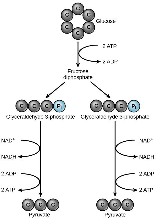
Simplified overview of glycolysis

Glycolysis begins with one 6-carbon glucose molecule and ends with two 3-carbon sugars, called pyruvate. In addition, two ATP molecules are produced and the coenzyme NAD+ is reduced to NADH. Reduced, high energy NADH molecules are produced to play their role as electrons (and H+) carriers (Figure 12.2.1.).

A graphic shows glucose at the top with an arrow pointing down to fructose diphosphate, which then splits into two glyceraldehyde 3-phosphate molecules. Each of these forms one NADH and two ATP molecules in the process of each becoming a pyruvate molecule.
Figure 12.2.1: Simplified glycolysis showing investment, cleavage and harvest.

Details of Glycolysis

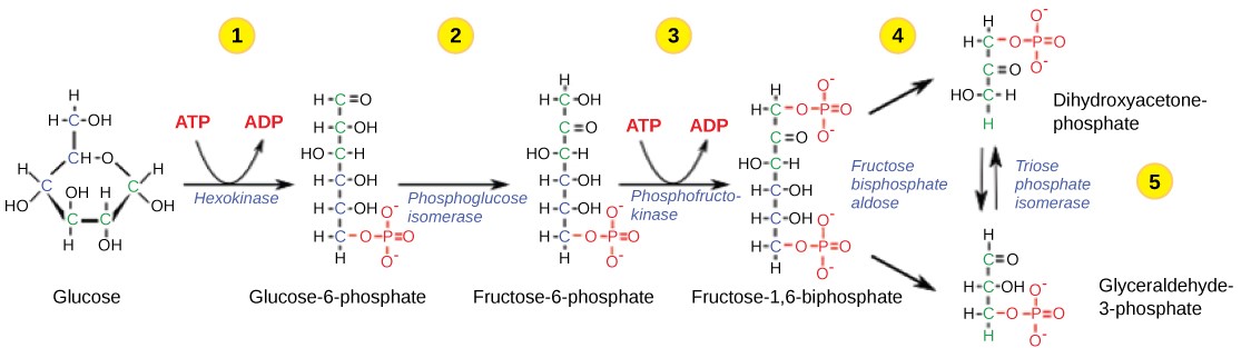
Glycolysis is a ten-step metabolic pathway

  • The first five steps (1-5) are the ATP-investment phases:  ATP is used to convert glucose to a three-carbon intermediate sugar, glyceraldehyde phosphate (Figure 12.2.2.).
  • The next five steps (6-10) are production phases; enzyme-catalyzed reactions and atom rearrangements  result in the final products – ATP, reduced coenzyme NADH and pyruvate (Figure 12.2.3.).
This illustration shows the steps in the first half of glycolysis. In step one, the enzyme hexokinase uses one A T P molecule in the phosphorylation of glucose. In step two, glucose dash 6 dash phosphate is rearranged to form fructose dash 6 dash phosphate by phosphoglucose isomerase. In step three, phosphofructokinase uses a second A T P molecule in the phosphorylation of the substrate, forming fructose dash 1, 6 dash bisphosphate. The enzyme fructose bisphosphate aldose splits the substrate into two, forming glyceraldeyde dash 3 dash phosphate and dihydroxyacetone-phosphate. In step 4, triose phosphate isomerase converts the dihydroxyacetone-phosphate into glyceraldehyde dash 3 dash phosphate.
Figure 12.2.2: The first half of glycolysis uses two ATP molecules in the phosphorylation of glucose, which is then split into two three-carbon molecules.
This illustration shows the steps in the second half of glycolysis. In step six, the enzyme glyceraldehydes dash 3 dash phosphate dehydrogenase produces one N A D H molecule and forms 1 3 dash bisphosphoglycerate. In step seven, the enzyme phosphoglycerate kinase removes a phosphate group from the substrate, forming one A T P molecule and 3 dash phosphoglycerate. In step eight, the enzyme phosphoglycerate mutase rearranges the substrate to form 2 dash phosphoglycerate. In step nine, the enzyme enolase rearranges the substrate to form phosphoenolpyruvate. In step ten, a phosphate group is removed from the substrate, forming one A T P molecule and pyruvate.
Figure 12.2.3: The second half of glycolysis involves phosphorylation without ATP investment (step 6) and produces two NADH and four ATP molecules per glucose.

Glycolysis is an anaerobic process: the reactions can occur independent of the present of oxygen. If Oxygen levels in the cells are LOW, pyruvate chemical reactions will result in the formation of lactic acid. While in the presence of sufficient oxygen, pyruvate continues to Phase 2 (Citric acid cycle).

Mitochondrion structure

The next two phases take place in different locations of the mitochondrion.  Take a moment to review its structure, paying close attention to the matrix, inner membrane, and intermembrane space.  You can refer back to the diagram as you read the next sections (Figure 12.2.4.).

           
This illustration shows the structure of a mitochondrion, which has an outer membrane and an inner membrane. The inner membrane has many folds, called cristae. The space between the outer membrane and the inner membrane is called the intermembrane space, and the central space of the mitochondrion is called the matrix. A T P synthase enzymes and the electron transport chain are located in the inner membrane.
Figure 12.2.4: Oxidative phosphorylation takes place in mitochondria. (Credit: modification of work by Mariana Ruiz Villareal)

License and attributions:

  • Biology, Second edition, 2018, Clark, M.A. et al. License: CC BY 4.0. Located at https://openstax.org/books/biology-2e/pages/7-2-glycolysis
  • Concepts of Biology, 2013, Fowler, S. et al. License: CC BY 4.0. Located at https://openstax.org/books/concepts-biology/pages/4-2-glycolysis

License

Icon for the Creative Commons Attribution 4.0 International License

BIO130: Introduction to Physiology Copyright © 2024 by Dinor Dhanabala; Sandra Fraley; and Gordon Lake is licensed under a Creative Commons Attribution 4.0 International License, except where otherwise noted.

Share This Book