"

Chapter 8: DNA Replication and Protein Synthesis

8.2. Protein Synthesis

Learning Objectives

By the end of this section, you will be able to:

  • Explain how the genetic code stored within DNA determines the protein that will form.
  • Describe the process of transcription.
  • Describe the process of translation.
  • Describe the roles of mRNA, tRNA and rRNA in protein synthesis.
  • Describe the function of ribosomes.
  • Explain the importance of the genetic triplet code.
DNA contains the information necessary for the cell to build a very important type of molecule: the protein. Most structural components of the cell are made up, at least in part, by proteins and virtually all the functions that a cell carries out are completed with the help of proteins. We’ve reviewed the many important roles of proteins as enzymes, oxygen carriers (hemoglobin), transporters across the cell membrane, cell and tissue structural components (collagen, elastin).

Protein synthesis

  • The process occurs in two different cell locations (Figure 8.2.1.)
    • Transcription occurs in the cell nucleus where mRNA is synthesized from a DNA template. After undergoing modification, mRNA moves outside the nucleus for translation.
    • Translation occur in the cytosol on ribosomes where mRNA’s coded information is translated to the correct amino acid sequence for the new protein.
  • A common strategy throughout protein synthesis is complementary base pairing.
This figure shows a schematic of a cell where transcription from DNA to mRNA takes place inside the nucleus and translation from mRNA to protein takes place in the cytoplasm.
Figure 8.2.1: From DNA to Protein.

Transcription:  DNA to mRNA 

DNA contains the genes. A gene is the functional segment of DNA (a nitrogenous base sequence) that specifies the order in which amino acids are to be placed in the peptide (or polypeptide) chain being synthesized. Activation of this DNA segment is called gene expression and transforms the information coded in a gene to a final gene product. Gene expression ultimately dictates the structure and function of a cell by determining which proteins are made.

In transcription, one DNA strand serves as the template for the synthesis of mRNA with base pairs that complement those of DNA (Figure 8.2.2.).  mRNA carries the genetic code for a protein’s amino acids as units of three nitrogenous bases. These are called the mRNA codons. For example, if TAC (Thymine-Adenine-Cytosine) is the nitrogenous base sequence on a segment of the DNA template, the mRNA codon will have AUG (Adenine-Uracil-Guanine). The newly synthesized mRNA strand undergoes post-transcriptional modification and the resulting so-called mature mRNA leaves the nucleus destined for the ribosome. The mature mRNA is a single linear strand of nucleotides which bases complementary to those of the DNA gene.

This diagram shows the translation of RNA into proteins. A DNA template strand is shown to become an RNA strand through transcription. Then the RNA strand undergoes translation and becomes proteins.
Figure 8.2.2: The Genetic Code DNA holds all of the genetic information necessary to build a cell’s proteins. The nucleotide sequence of a gene is ultimately translated into an amino acid sequence of the gene’s corresponding protein.

Translation:  mRNA to Protein

Translation occurs outside the nucleus on a ribosome which is organized in large and small subunits (Figure 8.2.3.). mRNA binds to the ribosomal small subunit and tRNA binds to its large subunit. Ribosomes are found either free in the cytosol or attached to an organelle called the endoplasmic reticulum (discussed in a later chapter).

The ribosome consists of a small subunit and a large subunit, which is about three times as big as the small one. The large subunit sits on top of the small one. A chain of m R N A threads between the large and small subunits. A protein chain extends from the top of the large subunit.
Figure 8.2.3: A large subunit (top) and a small subunit (bottom) comprise ribosomes. During protein synthesis, ribosomes assemble amino acids into proteins.

Translation requires mRNA, ribosomal RNA (rRNA) and transfer RNA (tRNA).

A ribosome is a macromolecule composed of many polypeptides and ribosomal RNA (rRNA). rRNA is a specialized enzyme-like catalyst called a ribozyme which functions to catalyze the formation the peptide bonds between amino acids during polypeptide formation. Ribosomes (rRNA) are synthesized in a specialized region of the cell nucleus called the nucleolus and then transported out to exist either free in the cytosol or attached to certain cell organelles. (Details of cell organization is discussed in a later chapter.)

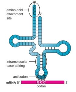
The tRNA molecule has a clover-leaf shape and two binding sites (Figure 8.2.4.).  One end of the tRNA carries an amino acid and the other end contains a triplet nucleotide sequence (the anticodon). Twenty amino acids are involved in making proteins and each amino acid has its own carrier tRNA carrier.

shows a single strand folded into a cross shape with intramolecular base pairing. The 3’ end at the top is labeled amino acid attachment site and has the sequence ACC. The 5’ end is also at the top. At the base of the cross is a three letter grouping called anticodon. This is complementary to a three letter set on the mRNA called a codon.
Figure 8.2.4: tRNA structure. One end carries a specific amino acid and the other end contains the anticodon (a triplet nucleotide sequence) that can bind only an mRNA codon.
In translation, the correct order of amino acids in the protein being synthesized is ensured by RNA anticodon-mRNA codon binding. mRNA codons containing the genetic code for specific amino acids interact with the anticodons on tRNA, the carrier of the amino acids. For example, mRNA codon GCC binds a tRNA anticodon CGG, its complementary base. The tRNA with this anticodon carries the amino acid Alanine (Ala) (Figures 8.2.4. & 8.2.5.).
Translation begins when a tRNA anticodon recognizes a codon AUG on mRNA. The resulting codon-anticodon binding results in Met being the first amino acid in the peptide chain (Figure 8.2.5.). The large ribosomal subunit joins the small subunit, and a second tRNA recognizes the next mRNA codon in ‘reading frame’ on the mRNA chain. Specific amino acids continue to be brought in by anticodon-codon recognition, ribozymes catalyze peptide bond formation in the growing amino chain, until a STOP signal codon is reached and the peptide chain dissociates from the ribosome.  
Illustration shows the steps of protein synthesis. First, the initiator tRNA recognizes the sequence AUG on an mRNA that is associated with the small ribosomal subunit. The large subunit then joins the complex. Next, a second tRNA is recruited at the A site. A peptide bond is formed between the first amino acid, which is at the P site, and the second amino acid, which is at the A site. The mRNA then shifts and the first tRNA is moved to the E site, where it dissociates from the ribosome. Another tRNA binds at the A site, and the process is repeated.
Figure 8.2.5: Formation of the peptide chain during translation.
The ‘dictionary’ of triplet genetic codes for amino acids is known (Figure 8.2.6.). Several points should be noted about this dictionary. 1) there is one START codon; 2) there are three STOP codons; 3) The dictionary contains synonyms (has redundancy). In other words, several codons may specify the same amino acid. For example, four different codons specify the amino acid Serine (Ser). One theory explaining the persistence of redundancy in the genetic code is that it offers protection against DNA mutations (nucleotide changes) that might render a protein nonfunctional due to shape changes.
Figure shows all 64 codons. Sixty-two of these code for amino acids, and three are stop codons.
Figure 8.2.6: The Genetic Code

License and attributions:

  • Anatomy and Physiology, Second edition, 2022, Betts, J.G. et al. License: CC BY 4.0. Located at https://openstax.org/books/anatomy-and-physiology-2e/pages/3-4-protein-synthesis
  • Concepts of Biology, 2013, Fowler, S. et al. License: CC BY 4.0. Located at https://openstax.org/books/concepts-biology/pages/9-4-translation
  • Microbiology, 2016, Parker, N. et al. License: CC BY 4.0. Located at https://openstax.org/books/microbiology/pages/11-4-protein-synthesis-translation
  • Biology, Second edition, 2018, Clark, M.A. et al. License: CC BY 4.0. Located at https://openstax.org/books/biology-2e/pages/4-3-eukaryotic-cells

License

Icon for the Creative Commons Attribution 4.0 International License

BIO130: Introduction to Physiology Copyright © 2024 by Dinor Dhanabala; Sandra Fraley; and Gordon Lake is licensed under a Creative Commons Attribution 4.0 International License, except where otherwise noted.

Share This Book